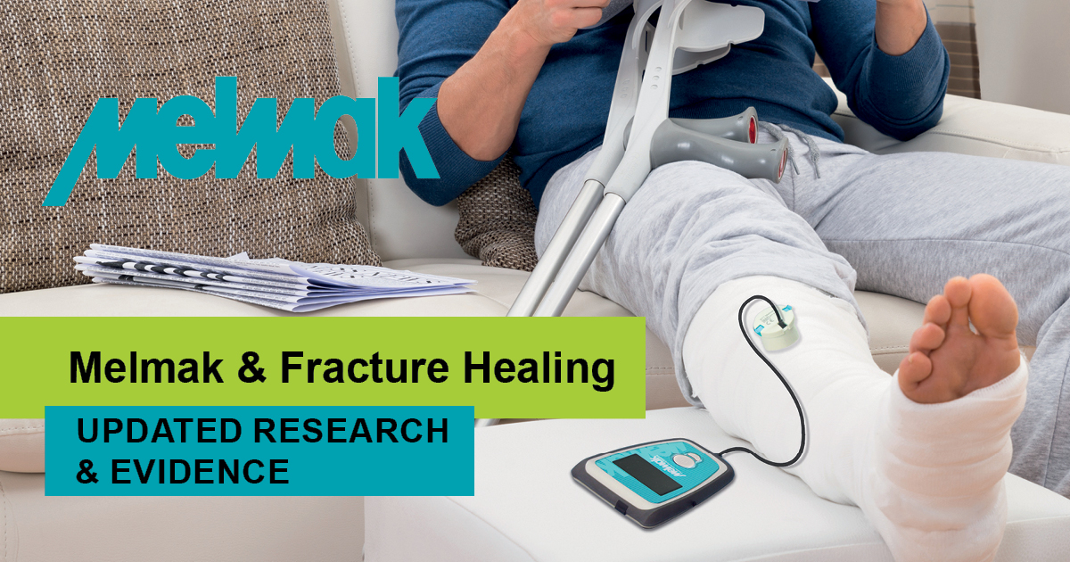
Of the estimated 156,000 fractures resulting from poor bone health within a year in Australia1, approximately 5-10% demonstrate delayed healing or non-union2, a figure that is set to increase as the survival rate for people with serious injuries improves.3 These delayed or non-healing fractures put a strain on both people, their families and the health care system, often leading to long-term complications that have a high monetary, psychological and physiological cost.
A variety of interventions including surgeries and bone grafts are proposed, despite carrying significant risks and lengthy recovery times.4,5 Low-intensity pulsed ultrasound (LIPUS) is one treatment that has gained significant popularity and commendation over the past decade, despite dating back 30 years since the first level one clinical trial demonstrated that LIPUS could accelerate fracture repair.6
The Melmak is a leading LIPUS device that is used by practitioners and clinics worldwide, backed by a rapidly growing body of evidence supporting the use of LIPUS in treating difficult-to-heal fractures. This article has summarised what you need to know about the device, the evidence supporting LIPUS, and how it compares to alternative treatments for non-union fractures.
Impaired Fracture Healing
Fracture healing is a complex process. Clinically, it requires adequate reduction of the displaced fracture and stabilisation.7 A delayed union is defined as healing that is not completed by three months from sustaining the fracture, and of those classified as delayed unions, some will still remain unhealed nine months post-fracture and are thus classified as non-unions.8
During the three stages of the fracture healing process,9 various cell types must interact and deliver appropriate levels of inflammatory and bioactive molecules. Despite the complexity of the healing process, most fractures heal without a problem. However, several risk factors are associated with impaired healing which include:10
● Presence of systemic disease, such as diabetes11 or advanced aging12,13
● External chemical factors, such as alcohol abuse14 or smoking15
● Prescribed medications like chronic steroid use or chemotherapeutics16
These risk factors provide an ongoing clinical challenge for the treatment of fractures in our aging, comorbid population. Several biophysical agents have been proposed and analysed as therapeutic adjuncts to address this impaired bone healing process.17
Treating Non-Union & Delayed Union Fractures
While advances in both operative and non-operative care of fractures continue to improve patient outcomes, under the best of circumstances, recovery times from the treatment itself still often extend from weeks to months.18 Slow healing or non-union fractures can have substantial economic consequences when one considers re-operation, secondary surgical procedures, and extended physical therapy in treating these fractures.19
Many treatment options are used in an attempt to accelerate fracture healing and prevent delayed healing and non-unions. These include bone grafting, alteration of the mechanical stiffness of the fixation devices, electromagnetic fields, high-frequency low-magnitude mechanical stimuli, and ultrasound.
Disadvantages of bone grafting and implantation of electrical stimulators include that these procedures must be performed in the operating theatre and often require hospital admission. The result is added morbidity to the patient and added cost to the health care system.20
Melmak LIPUS Evidence
A continually growing body of evidence supports the use of low-intensity pulsed ultrasound for the treatment of both fresh fractures21,22 and non-unions.23,24,25 An early meta-analysis of LIPUS found an average difference in healing time across 6 studies of 64 days between the treatment and control groups.26
While results on healing time improvements across studies vary, it is clear that research shows consistently that LIPUS treatment significantly reduces healing time in a range of fractures and settings.
It has been demonstrated that LIPUS has positive effects at each phase of the fracture healing process.27,28 Early research found that the non-union effectiveness rate for patients using one of the pulsed electromagnetic systems daily was 80%29. LIPUS therapy on tibial and radial shaft fractures showed that the healing time for tibial fractures was reduced by an average of 34% in testing groups compared to the control group. Similarly, healing time for distal radial fractures was reduced by 43% in testing groups compared to the control group.
LIPUS treatment also resulted in a trend toward a reduced incidence of tibial delayed union.30 The pooled results from the 3 studies showed that the time to healing was significantly shorter in the groups receiving low-intensity pulsed ultrasound treatment than in the control groups.31
There is also evidence that LIPUS can support healthy blood supply to fractured areas. Blood supply is a central variable for prompt and successful healing, and it is widely recognised that medical comorbidities that contribute to impaired blood flow will inhibit the ability of a fracture to heal. Research examining the volume density of blood vessels found that it was significantly increased in the LIPUS-treated delayed-union fractures group compared to placebo control group. LIPUS did not change blood vessel number, but significantly increased blood vessel size.32 Another study showed LIPUS to significantly improve short-term microcirculation when applied to the foot.33
How LIPUS works
Studies show that LIPUS causes the stimulation of a biological response within the cells, producing bioactive molecules. It is these molecules that play a key role in fracture healing.34
When defining ‘low intensity’ for LIPUS, we refer to an intensity of 30 mW/cm2 SATA (spatial average–temporal average) and a power of 117 mW delivered via an unfocussed ultrasound transducer. The ultrasound carrier frequency is 1.5 MHz, pulsed at 1 kHz.35 The image below shows the pulse duration, off time and intensity of LIPUS.
[ux_image_box img=”37330″]
Image credit: Hadjiargyrou M, McLeod K, Ryaby JP, Rubin C. Enhancement of fracture healing by low intensity ultrasound. Clin Orthop 1998;355(Suppl):s216-29.
[/ux_image_box]
As ultrasound waves pass through tissues, absorption of the waves is proportionate to the density of the tissue. This may explain how it can be efficiently targeted to the fracture gap, since bone has a significantly higher density than the surrounding soft tissues.36 Evidence states that a molecular response causes a number of genes to be expressed in response to LIPUS, and the products of the expression of those genes plays a key role in callus formation and stability37,38,39,40. One particular gene, aggrecan, which has increased expression after LIPUS use, is correlated with an increase in torsional strength of the calluses treated with ultrasound.41
Advise for Use
When considering low-intensity pulsed ultrasound, it is important to note that not all ultrasound machines will be appropriate. High-intensity continuous-wave ultrasound has been found to be deleterious to fracture healing, unlike the low-intensity pulsed ultrasound discussed previously.42,43,44,45,46
The differences between conventional ultrasound therapies used in general rehabilitation and the LIPUS therapy are the duration of the treatment and the wave parameters. Conventional ultrasound has been used in physiotherapy for decades for improved joint mobility and pain reduction in traumas47, but mostly due to the thermal effect48. In contrast, LIPUS in the therapy of bone healing is usually applied daily and for longer durations.
Generally, a treatment time of 20-30 minutes per day will support healing. Patients who used the device for 30 minutes per day were found to have a nonunion heal rate of 60.7%49, while 20 minute treatment times have found a 38% acceleration of fresh fractures.50,51
As LIPUS has a much lower intensity than the ultrasound generated from most regular clinical machines, the Melmak LIPUS device is portable and easy-to-use, facilitating the implementation of home-based rehabilitation interventions, increasing patient compliance, and optimising success rates.
Melmak: An Asset to Patients, Practitioners & Clinics
Amongst the modalities available to enhance fracture healing, Melmak is safe, practical, and effective. It can easily be administered without pain, by the patient in their home, without the need for hospital admission, anaesthesia, additional surgical procedures, and lengthy recovery times from the treatment itself. Additionally, Melmak is providing clinics with an excellent return on their investment , and is proving to be the perfect fit for clinics that are offering injury rehab or sports medicine services .
For more information about the Melmak LIPUS device, or to purchase this exclusive device, please contact RehaCare Customer Service on 1300 653 522 or at info@rehacare.com.au.
[divider width=”medium”]
[1] https://www.garvan.org.au/news-events/files/know-your-bones-consumer-report-2018.pdf
[2] Einhorn TA. Enhancement of fracture-healing. J Bone Joint Surg Am 1995; 77:940-56.
[3] https://jamanetwork.com/journals/jamasurgery/fullarticle/2547685
[4] Duarte LR. The stimulation of bone growth by ultrasound. Arch Orthop Trauma Surg 1983;101:153-9.
[5] Hadjiargyrou M, McLeod K, Ryaby JP, Rubin C. Enhancement of fracture healing by low intensity ultrasound. Clin Orthop 1998;355(Suppl):s216-29.
[6] 10.1016/j.ultras.2016.03.016
[7] 10.1016/j.ultras.2016.03.016
[8] Hadjiargyrou M, McLeod K, Ryaby JP, Rubin C. Enhancement of fracture healing by low intensity ultrasound. Clin Orthop 1998;355S:S216—29.
[9] https://www-sciencedirect-com.ezproxy.massey.ac.nz/science/article/pii/S002013830800048X?via%3Dihub
[10] 10.1016/j.ultras.2016.03.016
[11] L. Oei, F. Rivadeneira, M.C. Zillikens, E.H. Oei, Diabetes, diabetic complications, and fracture risk, Curr. Osteoporos. Rep. 13 (2) (2015) 106–115.
[12] J.D. Heckman, J. Sarasohn-Kahn, The economics of treating tibia fractures. The cost of delayed unions, Bull. Hosp. Joint Dis. 56 (1) (1997) 63–72.
[13] A.A. Naik, C. Xie, M.J. Zuscik, P. Kingsley, E.M. Schwartz, H. Awad, R. Guldberg, H. Drissi, J.E. Puzas, B. Boyce, X. Zhang, R.J. O’Keefe, Reduced COX-2 expression in aged mice is associated with impaired fracture healing, J. Bone Miner. Res. 24 (2) (2009) 251–264.
[14] M.S. Gaston, A.H. Simpson, Inhibition of fracture healing, J. Bone Joint Surg. Br. 89 (12) (2007) 1553–1560.
[15] J.A. Bishop, A.A. Palanca, M.J. Bellino, D.W. Lowenberg, Assessment of compromised fracture healing, J. Am. Acad. Orthop. Surg. 20 (5) (2012) 273– 282.
[16] E.M. Kagel, T.A. Einhorn, Alterations of fracture healing in the diabetic condition, Iowa Orthop. J. 16 (1996) 147–152.
[17] 10.1016/j.ultras.2016.03.016
[18] https://www-sciencedirect-com.ezproxy.massey.ac.nz/science/article/pii/S002013830800048X?via%3Dihub
[19] Heckman JD, Sarasohn-Kahn J. The economics of treating tibia fractures: the cost of delayed unions. Bull Hosp Joint Diseases 1997;56(1):63—72.
[20] https://www-sciencedirect-com.ezproxy.massey.ac.nz/science/article/pii/S002013830800048X?via%3Dihub
[21] J.D. Heckman, J.P. Ryaby, J. McCabe, J.J. Frey, R.F. Kilcoyne, Acceleration of tibial fracture-healing by non-invasive, low-intensity pulsed ultrasound, J. Bone Joint Surg. Am. 76A (1) (1994) 26–34.
[22] T.K. Kristiansen, J.P. Ryaby, J. McCabe, J.J. Frey, L.R. Roe, Accelerated healing of distal radial fractures with the use of specific, low-intensity ultrasound, J. Bone Joint Surg. Am. 79A (7) (1997) 961–973.
[23] P.A. Nolte, A. van der Krans, P. Patka, I.M.C. Janssen, J.P. Ryaby, G.H.R. Albers, Low-intensity pulsed ultrasound in the treatment of nonunions, J. Trauma 51 (4) (2001) 693–703.
[24] D. Gebauer, E. Mayr, E. Orthner, J.P. Ryaby, Low-intensity pulsed ultrasound: effects on nonunions, Ultrasound Med. Biol. 31 (10) (2005) 1391–1402.
[25] X. Roussignol, C. Currey, F. Duparc, F. Dujardin, Indications and results for the ExogenTM ultrasound system in the management of non-union: a 59-case pilot study, Orthop. Traumatol. Surg. Res. 98 (2) (2012) 206–213.
[26] https://www.cmaj.ca/content/166/4/437.long
[27] Y. Azuma, M. Ito, Y. Harada, H. Takagi, T. Ohta, S. Jingushi, Low-intensity pulsed ultrasound accelerates rat femoral fracture healing by acting on the various cellular reactions in the fracture callus, J. BoneMiner. Res. 16 (4) (2001) 671–680.
[28] https://www-sciencedirect-com.ezproxy.massey.ac.nz/science/article/pii/S002013830800048X?via%3Dihub
[29] D.E. Garland, B. Moses, W. Salyer, Long-term follow-up of fracture nonunions treated with PEMFs, Contemp. Orthoped. 22 (3) (1991) 295–302.
[30] https://www.cmaj.ca/content/166/4/437.long
[31] https://www.cmaj.ca/content/166/4/437.long
[32] https://pubmed.ncbi.nlm.nih.gov/29436706/
[33] https://pubmed.ncbi.nlm.nih.gov/28735428/
[34] 10.1016/j.ultras.2016.03.016
[35] 10.1016/j.ultras.2016.03.016
[36] Hadjiargyrou M, McLeod K, Ryaby JP, Rubin C. Enhancement of fracture healing by low intensity ultrasound. Clin Orthop 1998;355S:S216—29.
[37] Hadjiargyrou M, McLeod K, Ryaby JP, Rubin C. Enhancement of fracture healing by low intensity ultrasound. Clin Orthop 1998;355S:S216—29.
[38] Naruse K, Miyauchi A, Itoman M, Mikuni-Takagaki Y. Distinct anabolic response of osteoblast to low-intensity pulsed ultrasound. J Bone Miner Res 2003;18(2):360—9.
[39] Wu C-C, Lewallen DG, Bolander ME, et al. Exposure to low intensity ultrasound stimulates aggrecan gene expression by cultured chondrocytes. Trans Orthop Res Soc 1996;21: 622.
[40] Yang K-H, Parvizi J, Wang S-J, et al. Exposure to low density ultrasound increases aggrecan gene expression in a rat femur fracture model. J Orthop Res 1996;14:802—9.
[41] Yang K-H, Parvizi J, Wang S-J, et al. Exposure to low density ultrasound increases aggrecan gene expression in a rat femur fracture model. J Orthop Res 1996;14:802—9.
[42] Hadjiargyrou M, McLeod K, Ryaby JP, Rubin C. Enhancement of fracture healing by low intensity ultrasound. Clin Orthop 1998;355S:S216—29.
[43] Malizos KN, Hantes ME, Protopappas V, Papachristos A. Low-intensity pulsed ultrasound for bone healing: an overview. Injury 2006;37S:S56—62.
[44] Reher P, Elbeshir el-NI, Harvey W, et al. The stimulation of bone formation in vitro by therapeutic ultrasound. Ultrasound Med Biol 1997;23:1251—8.
[45] Tsai CL, Chang WH, Liu TK. Preliminary studies of duration and intensity of ultrasonic treatments on fracture repair. Chin J Physiol 1992;35:21—6.
[46] Wang SJ, Lewallen DG, Bolander ME, et al. Low intensity ultrasound treatment increases strength in a rat femoral fracture model. J Orthop Res 1994;12:40—7.
[47] J. D. Heckman et al., “Acceleration of tibial fracture-healing by non-invasive, low-intensity pulsed ultrasound,” J. Bone Joint Surg. Am., vol. 76, no. 1, pp. 26–34, Jan. 1994.
[48] T. Watson, “Ultrasound in contemporary physiotherapy practice,” Ultrasonics, vol. 48, no. 4, pp. 321–329, Aug. 2008.
[49] http://www.djoglobal.com/products/cmf/cmf-ol1000
[50] J.D. Heckman, J.P. Ryaby, J. McCabe, J.J. Frey, R.F. Kilcoyne, Acceleration of tibial fracture-healing by non-invasive, low-intensity pulsed ultrasound, J. Bone Joint Surg. Am. 76A (1) (1994) 26–34.
[51] T.K. Kristiansen, J.P. Ryaby, J. McCabe, J.J. Frey, L.R. Roe, Accelerated healing of distal radial fractures with the use of specific, low-intensity ultrasound, J. Bone Joint Surg. Am. 79A (7) (1997) 961–973.


